The Blow Fly
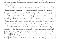
The Blow Fly (Musca vomitoria) is an insect belonging to the order Diptera, a large and important tribe, the leading characters of which are the possession of two wings (hence the name), and a mouth arranged for sucking.
Insects are articulate (jointed) animals in which the external covering or integument serves the same purpose as the bony skeleton of vertebrate animals.
From vertebrate animals they differ perhaps more greatly still in development. There are usually three well defined periods in the life of an insect, known as the larval, pupae, & imaginal states.
In the larval condition growth is exceedingly rapid, the hard integument being usually cast off periodically & replaced by new layers formed beneath it. When fully grown the insect generally takes a different form, known as a pupae or chrysalis, when inactive, and a nymph, when more or less active.
After a time, the pupa case bursts, or the nymph sheds its skin, & the perfect insect comes forth, rapidly acquiring its full powers, when growth & development cease. In some cases the larva, pupa & imago or perfect insect closely resemble each other, in others, as in the Blow Fly, the differences between the larva & imago is so great that doubt might be thrown upon their being the same individual.
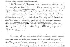
The larvae of Diptera are commonly known as maggots or gentles. In the process of development of the Blow Fly such radical changes take place that it is asserted on the highest authority that not one structure is present in the fly as it exists in the maggot. Every portion of the larva, except the brain & perhaps a portion of the alimentary canal undergoes rapid degeneration, the fly being formed within the pupa skin by a process of redevelopment.
The time at our disposal this evening will permit me to notice only the more important organs of the fly: a detailed description of all would require many months preparation & would furnish matter for several papers. I propose therefore to confine myself to the Legs, Wings, Organs of Respiration, Proboscis, & Eyes.
The legs consist of four parts – the Coxa or hip, uniting the limb to the Thorax, the Femur or thigh, the Tibia or leg & the Tarsus or foot. The Tarsus consists of 5 pieces the last bearing a pair of pads termed Pulirlli & a pair of hooks just above the Pads. Both hooks & pads are connected toa fine tendon running trough the Tarsus & Tibia & ending in a muscle in the interior of the Femur.
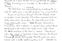
The Foot Pads are very curious & interesting, enabling the insect to walk upon a smooth surface & in any position.
It was at one time thought that the Pulirlli acted as suckers, more recently it has been supposed to be due to exhaustion of air from the disc like end of the hairs clothing the pads. Careful observations have, however, shown that adhesion is caused by a viscid fluid exuding from the extremities of the hairs with which the under surface of the pad is covered. “Sometimes when the insect is captured & held between the thumb & finger this viscid liquid exudes so rapidly that the pads are soon covered with a little glistening drop of it.” Collected on a glass slide the secretion quickly becomes solid. It is insoluble in water. When the leg is dissected from the body the whole contents of the Tarsus become solid very rapidly. “The feet of the smaller House Fly are the best to show the manner in which the viscid fluid exudes from the extremities of the trumpet shaped hairs as they are very large comparatively.” “The foot prints left upon glass by flies consist of rows of dots corresponding to these hairs” of which each pad has about 1200.
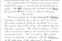
It is probable that the secretion never becomes solid whilst it remains in contact with the foot of the living insect. The pad is released from its hold by the Tarsus being curved so that it may be withdrawn obliquely.
The Wings
The wings consist of a double membrane extended over a framework of nervures the more important of which are placed on or near the anterior edge of the wing, the posterior edge being free from nervures. The whole membrane is covered with minute hairs those on the nervures being stronger & more prominent. The membrane forming the Wings is identical in structure with the Protoderm as the outer layers of the integument is termed.
Organs of Respiration
The respiratory organs of insects are arranged upon a plan differing widely from that of Vertebrate animals. In Insects provision is made for the aeration of the blood by the introduction of Air to all parts of the body through a ramifying system of tubes, called Trachea, from their cavity being kept open by a spiral Fibre, or more often by a series of rings which run into each other so as to appear spiral. These Tubes open outwardly by pores called Spiracles internally they undergo repeated subdivision until at last they become reduced to a diameter of nearly 100,00th of an inch permeating every organ. The Tracheal Tubes remind one forcibly of the spiral vessel of plants, this arrangement being the one calculated to secure the greatest strength with the smallest expenditure of material. They consist of two coats the spiral fibre being developed in the substance of the internal one & not being distinct from them as some observers have asserted.
The Spiracles or external openings are generally visible as a row of pores on either side of the Thorax & abdomen. They are usually protected by a vaturlar or an arborescent growth from the edges of the opening in the latter case forming a sort of sieve & probably serving as a filter for the air which passes through them. The manner in which the air is caused to circulate through the Trachea is not thoroughly known. From the rapidity with which Chloroform acts upon the insect there can be little doubt that the circulation is vey vigorous. It is believed that the pressure of the muscles of the Thorax acting unequally upon the main Trachea aided by the valves at the Spiracles mainly tends to propel the air through the smaller tubes.
In the male fly there are 10 spiracles on each side, in the female, two pairs less are present.
The circulation of the blood in the fly & in all insects takes place without the aid of true blood vessels. A single organ called the dorsal vessel runs the whole length of the back beginning near the apex of the abdomen & ending in the head. It is open at each end and acts as a heart pulsating regularly & pumping the circulating fluid from the hinder portion of the insect into the head whence it returns to the visara. This vessel makes about 180 pulsations in a minute.
The chief cause of the humming sound made by the insect seems to be the rapid vibration of the valves of the Trachea of the Thorax.
The Proboscis
This organ which is one of the most remarkable & complex structures to be found in the insect world is more or less familiar to all microscopists. No cabinet is complete without one of those beautiful preparations first introduced I believe by the late Mr. Topping. This mode of mounting although it reveals much of the delicate structure of the organ scarcely does justice to it.
To acquire a true conception of its wonderful adaptation so its intended purpose it must be seen with all its parts in their natural position & not distorted by pressure. Probably there is no better way of acquiring a knowledge of its structure & function than by watching the proboscis of a living fly in action. To do this the insect should be placed in a deep live box with a little thin syrup on the under surface of the glass cover & illuminated from above. The Proboscis may also be removed from the fly & soaked in Glycerin until it becomes transparent & then mounted in that fluid. Thus prepared, it forms a most beautiful object especially if examined with a Binocular under black ground illumination. Sections of the organ made with a lancet are also highly instructive showing well the minute internal structure.
The preparations of the Proboscis usually sold consist of the portion beyond the Pharynx & embracing the Maxillary Palpi, the Canula & the Lobes.
The Maxillary Palpi are comparatively large rod shaped organs, two in number, inserted on that part of the Proboscis just in front of the Pharynx called the Maxilla. They are covered with minute hairs & are also beset with numbers of larger bristle-like projections. “Whatever their function may be it is probably called into activity when the proboscis is retracted for they are turned back when it is exserted”; “they are possibly connected with the sense of Taste or Smell.
“The Canula or central portion is thick & flesh being filled with muscles. It is terminated by the lobes of the Proboscis & is deeply grooved on its dorsal surface, the groove forming the mouth & concealing the Tongue.”
The Lobes are two large fleshy processes terminating the Proboscis. When at rest these lobes are folded against each other but when opened about two thirds of their anterior surface forms a sucker of a some what oval shape divided into two parts by the fissure between them.
The anterior surface of the Lobes is furnished with a series of canals called false Trachea which open internally into the cavity between the lobes & so into the mouth: these form a fine strainer through which the insect is enabled to extract the fluid from the solid portion of the material upon which it feeds.
The Lobes are capable of further separation exposing a triangular opening surrounded by 50 to 60 rod-like teeth which are used for grinding hard substances such as sugar to facilitate their solution by the salivary secretion.
The channels of the false Trachea are kept open by incomplete rings which are forked at one extremity this forking occurring on either side alternately. The number of the channels is variable & not always the same in each lobe, there are generally 29 or 30 on either side. They open as follows. The 5 anterior channels unite and form a large trunk which runs along the upper border of each lobe and opens close to the upper margin of the triangular opening, the next 7 or 8 open separately each externally to a set of teeth & the remainder run into a pair of parallel channels in the middle of the disc.
The space between the parallel channels is covered with minute hairs. The arrangement of the false Trachea is exceedingly well shown in Mr. Topping’s preparation.
The Eyes
The Eyes of the Blow Fly consist of two compound clusters & three simple Eyes. As most interest attaches to the compound ones I shall more particularly describe them. They consist of an aggregation of organs each possessing the power of forming an image. These are termed Ocelli & together they form a nearly hemispherical cluster immovably fixed on either side of the head. Examined with a lens the surface is seen to be facetted, the facets being hexagonal in the centre of the cluster becoming more or less square towards the base. Each Facet is the Corneule of a separate Ocellus or little eye. The Corneule forms a double convex lens the diameter of each lens being about 1/1000 of an inch. Beneath the Corneule is a corresponding conical body partly separated by a layer of dark Pigment with a central perforation through which the rays of light which have passed through the lens gain access to the retina. The focus of each Corneule has been ascertained to be equivalent to the length of the pyramid behind it so that the image produced by the lens will fall upon the extremity of the filament of the optic nerve. The cones are separated from each other by pigment so that no rays except those which pass through the axis of each cone can reach the optic nerve. In each cluster of the Eyes of the common fly there are about 2000 Ocelli & it is probable that the combined action of the whole produces but a single image resembling that which we obtain with our two eyes.
In addition to the organs described I have slides of the Ovipositor & Antennae. The ovipositor in the blow fly is comparatively large & is well adapted for piercing flesh.
[The above lecture is hand written on eleven sheets of paper plus one blank sheet. Each sheet is watermarked with a series of eight equally spaced horizontal lines and measures 23 x 18 cm. It is unsigned or dated]
